Services


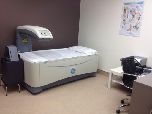
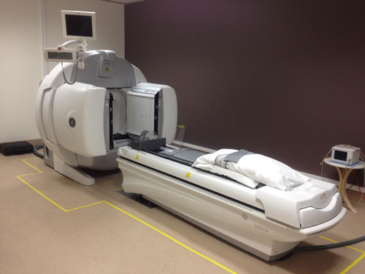
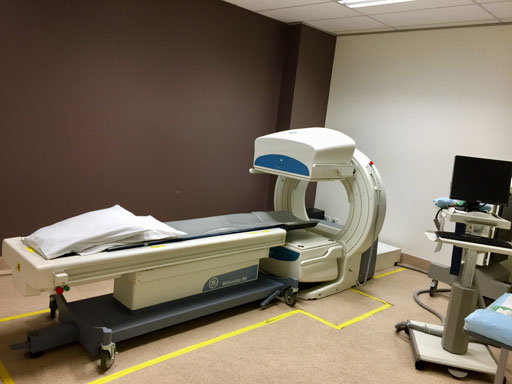
We have 2 GE Gamma cameras and a DEXA Densitometer allowing us to make detailed and accurate assessments.
We conduct a wide range of nuclear medicine scans from bone densitometry to lung scans. Feel free to view our extensive list of services.

Procedures we perform
A Bone Scan or Bone Scintigraphy is a nuclear medicine imaging of the bone. It is a “functional” imaging that is based on bone activity/metabolism (unlike X-Ray or CT-Scan which are “anatomical” imaging) and is very sensitive in diagnosing a number of bone conditions such as: cancer of the bone or metastasis, inflammatory arthritis, stress fractures (that may not be visible on X-ray or CT), and osteomyelitis (bone infection).
A Myocardial Perfusion scan (also referred to as MPI or MPS) is a nuclear medicine imaging performed with a stress test (either with exercise on a treadmill or pharmacologically with Persantin). MPI assess the blood flow of the coronary arteries that supply the myocardium (heart muscle) and hence can identify blocked or partially blocked coronary arteries. It can also identify previous myocardial infarction (scarring of the heart muscle after a heart attack) and assess the ‘pumping ‘action of the heart by calculating the left ventricular ejection fraction (LVEF). It is a non- invasive test and is has a high sensitivity and specificity comparable to CT coronary angiography.
A Gated Heart Pool Scan is a nuclear medicine imaging which involves labelling the red blood cells in your blood with a radiopharmaceutical and then measures the amount of blood in the heart that is pumped out with each heart beat. The images allow visualisation of the contraction of the heart muscles and computes the left ventricular ejection fraction (LVEF). It gives information on how well the heart is pumping.
A Lung Scan is a nuclear medicine imaging to diagnose pulmonary embolus (blood clots in the lung). It is important that treatment is commenced once the diagnosis is made to prevent new clots.
A Thyroid Scan is a nuclear medicine imaging that assess the function/activity of the thyroid gland. The thyroid gland controls the metabolism of the body and can become over or under active. It is commonly used to diagnose Grave’s disease ( over-active thyroid gland ) or Hashimoto’s thyroiditis ( under-active thyroid gland ) or subacute thyroiditis ( which is an inflammation of the thyroid gland ).
Thyroid scan is also used to assess if a thyroid nodule is toxic (“hot”) or non-functioning (“cold”). If it is non-functioning/cold, fine needle aspiration biopsy is needed to exclude the possibility of thyroid cancer.
A Parathyroid Scan is a nuclear medicine imaging to determine the function of the parathyroid glands which regulates calcium uptake in the body. It is commonly used in patients with hyperparathyroidism to look for a functioning parathyroid adenoma which can be surgically removed.
A Gastric Emptying Scan is a nuclear medicine imaging to determine how fast the stomach empties food into the small intestine after eating. There is no injection involved but instead the patient is given a radio-labelled egg sandwich and images are acquired at 15 minute intervals up to 2 -3 hours. It is used to look for patients with symptoms of gastroparesis such as early satiety, bloating sensation and nausea + vomiting.
A HIDA scan, also called Cholescintigraphy or Hepatobiliary Scintigraphy, is a nuclear medicine imaging used to assess gall bladder function. It is used to diagnose chronic acalculous cholecystitis or gallbladder dyskinesia which can present as obscure abdominal pain, especially when ultrasound, CT scan and gastroscopy findings are normal.
A Renal Scan is a nuclear medicine imaging scan that looks at the function of the kidneys such as the blood flow to the kidneys and urine flow from the kidneys to the bladder. It is used to investigate patients with hydronephrosis/obstruction and pyelonephritis/scarring following episodes of kidney infection.
Bone density, or bone mineral density, is the amount of bone mineral in bone tissue. Bone Densitometry is a test like an X-ray that quickly and accurately measures the density of bone. It is used primarily to detect osteopenia or osteoporosis, diseases in which the bone’s mineral and density are low and the risk of fractures is increased.
Osteoporosis ( “brittle” bones ) is a silent medical condition that can only be detected by DEXA . If you had a minimal trauma fracture, it is likely that you may have osteoporosis.
For more information please visit bonehealth.com.au
